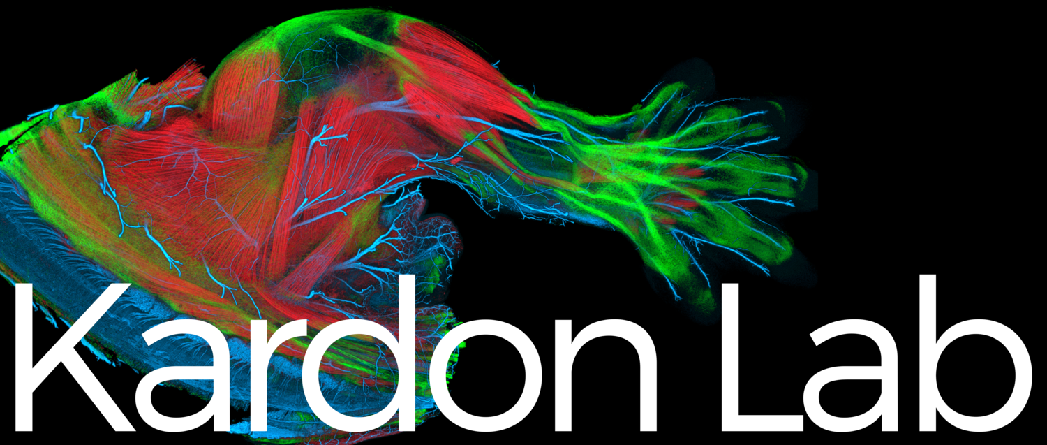
Our Research
Muscle Stem Cells in Development, Regeneration, and Homeostasis
Muscle development, growth, and regeneration take place throughout vertebrate life. Muscle stem cells are critical for all of these processes. Our research has concentrated on the developmental origin of muscle stem cells, their function, and the extrinsic signals that regulate them. We found that all muscle stem cells in the limb arise in the embryo from the somites, but there are related, although distinct populations of stem cells in the embryo, fetus, and adult (Kardon et al. 2002, Schienda et al. 2006, Hutcheson et al. 2009, Murphy et al. 2011). We established that muscle stem cells are critical for muscle development (Hutcheson et al. 2009) and adult muscle stem cells (known as satellite cells) are necessary and sufficient for muscle regeneration (Murphy et al. 2011). Adult muscle stem cells also contribute to muscle homeostasis, but their requirement is still not clear (Keefe et al. 2015). Most recently, we have reconstructed muscle regeneration in three dimensions (Collins, Shapiro et al. 2024). After a catastrophic injury (BaCl2), we show that myofiber regeneration occurs via two waves of fusion: the first wave of density-dependent fusion of myocytes re-establishes correctly oriented myofibers, while subsequent fusion of myocytes to myofibers enlarges regenerated myofibers. The residual basement membrane of damaged myofibers is critical for promoting the first wave of fusion and insures that regenerated myofibers are correctly oriented. A surprising finding of our research is that the centralized myonuclei are an enduring feature of regenerated myofibers, and we are currently testing whether these myofibers are biomechanically compromised.
Muscle Damage and Regeneration after Alphavirus Infection
Ross River (RRV) and Chikungunya (CHIKV) alphaviruses are emerging mosquito-borne pathogens that target the musculoskeletal system and a major health concern as they cause explosive outbreaks in humans. A major unknown in our understanding of RRV and CHIKV disease is how viral infection causes acute and chronic myalgia and myopathy, an important consequence of viral infection found in 90% of patients. In collaboration with Deborah Lenschow’s lab (Washington University, St. Louis) and with initial funding from a Pew Innovation Award, we are determining what functional, structural, cellular, and molecular changes in muscle result from RRV and CHIKV infection and testing whether infection of myogenic cells drives systemic acute and chronic disease.
Diaphragm: Development, CDH, and Evolution
The diaphragm is an essential mammalian skeletal muscle. It is vital for respiration and serves as a barrier between the abdomen the thorax (see our reviews - Merrell et al. 2013 and Kardon et al. 2017) . Unfortunately, defects in diaphragm development are the cause of congenital diaphragmatic hernias (CDHs), a common birth defect (1:300 births) that results in severe morbidity and 50% mortality. Using mouse genetics and ex vivo two photon imaging of the developing diaphragm, we definitively established that the pleuroperitoneal folds (PPFs), transient embryonic structures, and the muscle connective tissue fibroblasts derived from them critically regulate development of the diaphragm muscle. Furthermore, we showed that mutations in Gata4 in PPF fibroblasts are a cause of CDH (Merrell et al. 2015). More recently, we used whole-genome sequencing of CDH individuals and their parents to comprehensively reconstruct the genetic architecture of 4 CDH individuals and demonstrate that the etiology of CDH is heterogeneous and multifactorial (Bogenschutz et al. 2020). Also, we developed a novel PPF culture system to efficiently test the functional significance of candidate CDH genes (Bogenschutz et al. 2020). Finally, using conditional mutagenesis in mice, we have established that PPF expression of HGF is essential for recruitment and expansion of muscle in the developing diaphragm (Sefton et al. 2022). Currently, we are further exploring the important molecular interactions between PPF fibroblasts and muscle cells during normal diaphragm development and CDH.
Although the diaphragm is a functionally essential skeletal muscle in humans, the muscularized diaphragm is an evolutionary novelty that arose in and uniquely defines mammals. What genetic and developmental innovations led to this major mammalian innovation? The question of how morphologic novelties arise is a major question in evolutionary biology. To discover what developmental innovations led to the evolution of the muscularized diaphragm, we are comparing the development of birds, which represent an ancestral, non-muscularized state, with mice.
Development of the Limb Musculoskeletal System and Congenital Limb Abnormalities
Development of the musculoskeletal system requires the coordinated development of muscle, muscle connective tissue, tendon, and skeleton. How these tissues, derived from different embryonic sources, are patterned and assembled had been largely unknown. Using the limb as a model system, we have established that reciprocal interactions between muscle and tendon are essential for delimiting the major groups of limb muscles (Kardon 1998) and that the connective tissue regulates the pattern of individual muscles (Kardon et al. 2003) and muscle fiber type (Mathew et al. 2011). We have also found that specification of particular muscles is tightly linked to the specification of their bone origin and insertion sites, and mutations in the transcription factor Tbx3 leads to limb muscle and bone defects in Ulnar Mammary Syndrome (Colasanto et al. 2016).